Therapeutic ultrasound is one of the most widely used modalities in physiotherapy – a clinically validated technology that uses high-frequency sound waves to accelerate tissue healing, reduce pain, and restore mobility. Unlike medical ultrasound, therapeutic ultrasound is designed to interact with the body’s tissues at a cellular level, producing measurable biological effects that support recovery and pain management.
This guide covers everything you need to know: how the technology works, what conditions it treats, what clinical evidence supports its use, and what to look for in a professional therapeutic ultrasound device.
What Is Therapeutic Ultrasound?
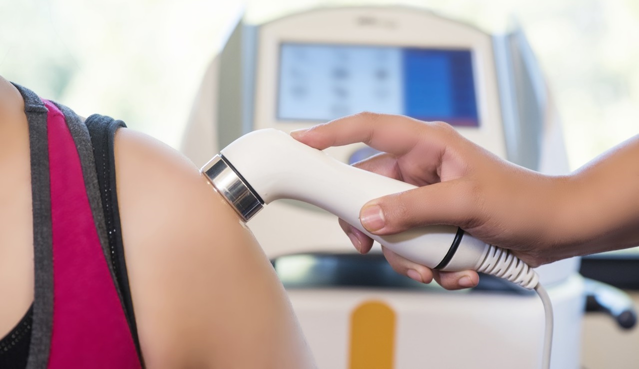
Therapeutic ultrasound is a non-invasive physiotherapy treatment that delivers acoustic energy deep into body tissues through a handheld transducer. The sound waves — typically operating between 1 and 3 megahertz (MHz) — penetrate the skin and underlying muscle layers, generating both thermal (heat-based) and non-thermal (mechanical) biological effects that promote healing and reduce inflammation.
Unlike diagnostic ultrasound, which uses sound to create images, therapeutic ultrasound is applied to treat musculoskeletal conditions, soft-tissue injuries, and chronic pain.
The Difference Between Therapeutic and Diagnostic Ultrasound
| Feature | Therapeutic Ultrasound | Diagnostic Ultrasound |
| Purpose | Tissue healing, pain relief | Medical imaging |
| Frequency | 1–3 MHz | 2–18 MHz |
| Output | Continuous or pulsed energy | Pulse-echo imaging |
| Duration | 5–10 minutes per area | Varies |
| Used by | Physiotherapists | Radiologists, sonographers |
A Brief History: How Ultrasound Entered Physiotherapy
Ultrasound’s therapeutic application dates to the 1940s, when researchers first observed that focused sound waves could produce localized biological effects in tissue. By the 1950s, physiotherapy clinics across Europe and North America had adopted ultrasound devices as a standard treatment tool. Decades of clinical refinement have led to the precise, programmable devices used in modern physiotherapy practice today.
How Does a Therapeutic Ultrasound Device Work?
A therapeutic ultrasound machine generates sound waves by passing an alternating electrical current through a piezoelectric crystal inside the transducer head. These waves travel through a coupling medium — typically a water-based gel — and penetrate the skin to reach target tissues.
The Piezoelectric Effect — Sound Waves from Electricity
The piezoelectric crystal at the core of every ultrasound transducer converts electrical current into mechanical energy, creating mechanical vibration at precise frequencies. These vibrations generate longitudinal sound waves that are beyond human hearing and propagate through soft tissue. The frequency and intensity can be adjusted by the clinician to target specific tissue depths and treatment goals.
The key principle: ultrasound energy enters body tissue, and its effects vary across different tissues depending on tissue type, stimulating cellular activity, improving circulation, and modulating pain signals.
Thermal Effects — Deep Tissue Heating Explained
When ultrasound waves are absorbed by tissue — particularly at the interface between soft tissue and bone — they generate localized deep heat. This thermal effect:
- Increases local blood flow and metabolism, which can help control pain by improving circulation and promoting healing
- Improves the extensibility of collagen-dense tissues (tendons, joint capsules)
- Reduces muscle spasm and stiffness, supporting pain control
- Enhances the effectiveness of concurrent stretching
Thermal effects are maximized using continuous wave ultrasound at 1–2 W/cm², whereas pulsed ultrasound is primarily non-thermal.
Non-Thermal (Mechanical) Effects — Cavitation and Acoustic Streaming
Even at low intensities that produce minimal heating, mechanical ultrasound creates slight vibrations in soft tissue and generates important mechanical effects:
- Stable cavitation: Microscopic gas bubbles in tissue fluids oscillate rhythmically, stimulating cell membrane activity and accelerating nutrient exchange
- Acoustic streaming: A unidirectional flow of fluid along cell membranes, increasing membrane permeability and supporting cellular repair, while helping remodel damaged tissue and scar tissue and reduce swelling and inflammation
- Microstreaming: At the cellular level, it stimulates fibroblast activity and protein synthesis, both critical for tissue regeneration and soft tissue healing
These non-thermal effects are maximized with pulsed ultrasound (typically 20% duty cycle), where the duty cycle creates rest periods between sound waves that limit heat while supporting healing.
1 MHz vs 3 MHz — Choosing the Right Frequency
The choice of frequency is one of the key ultrasound parameters and treatment parameters because it determines how deeply the ultrasound penetrates into tissue and where energy is absorbed. This is one of the most clinically important decisions a physiotherapist makes when setting up treatment.
| Parameter | 1 MHz | 3 MHz |
| Tissue penetration | Up to 5 cm | Up to 2 cm |
| Best for | deeper tissues | more superficial tissues |
| Heating rate | Slower, broader | Faster, more localised |
| Typical application | Hip, shoulder, lumbar | Ankle, wrist, knee, elbow |
Most professional therapeutic ultrasound devices offer selectable frequency, allowing clinicians to tailor treatment precisely to each patient’s anatomy and condition, especially when targeting superficial tissues. Frequency choice is also influenced by energy loss as sound passes through tissue, a process called attenuation in ultrasound physics.
What Conditions Does Ultrasound Therapy Treat?
Therapeutic ultrasound has a broad range of clinical applications across musculoskeletal and pain management settings. Below are the most well-supported conditions in both clinical practice and research literature.
Musculoskeletal and Soft Tissue Injuries
- Tendinopathy (including rotator cuff, Achilles, and patellar tendons)
- Muscle strains and tears (sub-acute phase)
- Ligament sprains
- Bursitis
- Myofascial trigger points
- Plantar fasciitis
Ultrasound therapy is often directed at collagen-rich structures such as ligaments and tendons because they absorb energy efficiently, which may produce a stronger therapeutic effect while promoting tissue healing through increased fibroblastic activity and protein synthesis.
Chronic Pain and Inflammation Management
Ultrasound therapy is one of the most frequently used physiotherapy modalities for Low Back Pain (LBP) and other chronic musculoskeletal conditions, where it may help reduce pain and muscle spasms. At 1–2 W/cm², therapeutic frequency stimulates local circulation, reduces pro-inflammatory cytokine activity, and promotes tissue remodeling over a course of treatment, with relief of muscle spasms among its broader therapeutic benefits.
Sports Injuries and Accelerated Recovery
Athletes benefit from ultrasound therapy’s ability to accelerate the sub-acute healing phase — reducing downtime after soft tissue injuries. Common applications include:
- Hamstring strains
- Ankle ligament sprains
- Tennis elbow (lateral epicondylalgia)
- Shin splints (medial tibial stress syndrome)
Post-Surgical Rehabilitation
Therapeutic ultrasound is frequently incorporated into post-surgical rehab protocols to reduce scar tissue formation, improve fascial mobility, and manage pain, and it may also support the healing process after surgery and the broader tissue healing process as patients return to function. Its use is typically part of clinical decision making, with modality selection based on timing and treatment goals, and modern guidelines recommend combining passive modalities with active rehabilitation such as therapeutic exercises alongside manual therapy and exercise-based rehabilitation.
| Condition | Treatment Goal | Typical Frequency | Evidence Level |
| Tendinopathy | Collagen remodelling, pain reduction | 1–3 MHz continuous | Moderate–High |
| Plantar fasciitis | Fascial mobility, pain relief | 1 MHz continuous | Moderate |
| Muscle strain (subacute) | Accelerate repair | 3 MHz pulsed | Moderate |
| Chronic LBP | Pain modulation, mobility | 1 MHz continuous | Moderate |
| Post-surgical scar | Scar remodelling | 3 MHz pulsed | Moderate |
| Bursitis | Inflammation reduction | 3 MHz pulsed | Low–Moderate |
Clinical Evidence — What the Research Shows
Key Studies on Ultrasound Therapy Effectiveness
The evidence base for therapeutic ultrasound spans decades and multiple systematic reviews. Key findings include:
- A 2015 Cochrane Review of ultrasound for non-specific chronic low back pain found that ultrasound was more effective than sham treatment for pain and function in the short term, though authors noted the need for larger trials.
- Research published in the Journal of Orthopedic & Sports Physical Therapy has supported ultrasound’s role in the management of tendinopathy, particularly when combined with eccentric loading exercises.
- Studies on phonophoresis — the use of ultrasound to enhance transdermal drug delivery — have shown effectiveness in delivering corticosteroids and anti-inflammatory agents into target tissues.
- Pulsed low-intensity ultrasound (LIPUS) has been extensively studied for fracture healing and fracture repair, with demonstrated efficacy in accelerating bone repair in delayed union fractures. In research settings, shockwave therapy and high intensity focused ultrasound are distinct ultrasound-based interventions studied for different clinical aims rather than routine soft-tissue physiotherapy use.
Note: Evidence quality varies across conditions. Physiotherapists integrate research findings with clinical expertise and individual patient presentation when designing treatment protocols.
What Physiotherapists Say About Ultrasound Treatment
Among physiotherapy practitioners, therapeutic ultrasound remains a valued modality for its versatility — offering both thermal and non-thermal treatment options, minimal side effects, and strong patient tolerance. Clinical experience consistently identifies its greatest value in the management of chronic tendinopathy and plantar fasciitis, and in trigger-point treatment when combined with manual therapy; in practice, physical therapists may also use ultrasound to improve range of motion and reduce joint stiffness when it is paired with manual therapy or exercise.
What to Expect During an Ultrasound Therapy Session
Before Your First Session — Assessment and Setup
Before beginning ultrasound therapy, your physiotherapist will:
- Conduct a full clinical assessment of your injury or condition.
- Confirm that no contraindications are present.
- Select the appropriate frequency, intensity, and treatment mode (continuous vs pulsed) as part of the treatment application, based on the target tissue and treatment goals.
- Apply ultrasound gel to the treatment area.
- Place the ultrasound head in direct contact with the patient’s skin when appropriate, keeping the planned treatment area to no more than about twice the effective radiating area (ERA) of the transducer head.
- Explain the sensations you may experience during treatment.
What Does Ultrasound Therapy Feel Like?
Most patients experience a mild warmth or gentle tingling sensation during treatment — particularly during continuous mode at therapeutic intensities. The treatment is generally pain-free.
If you feel sharp pain, excessive heat, or significant discomfort at any point, you should inform your physiotherapist immediately, as this may indicate incorrect application technique or an undetected contraindication.
Session Duration and the Number of Treatments Needed?
A typical ultrasound therapy session has a treatment duration of 5–10 minutes per treatment area. The full course of treatment depends on the condition:
- Acute injuries: 3–6 sessions, often combined with other modalities
- Sub-acute conditions: 6–10 sessions
- Chronic conditions: 10–15+ sessions, with regular reassessment
- Trigger point treatment: 1–5 sessions per trigger point area
Your physiotherapist will reassess your progress regularly and adjust the treatment plan based on your clinical response.
Combining Ultrasound with Other Physiotherapy Modalities
Therapeutic ultrasound is frequently combined with:
- Manual therapy — joint mobilization and soft tissue techniques
- Electrotherapy — TENS, interferential current, or laser therapy
- Exercise rehabilitation — the cornerstone of lasting outcomes
- Phonophoresis — topical medication delivery enhanced by ultrasound
- Dry needling or trigger point therapy — for myofascial conditions
This multimodal approach consistently outperforms ultrasound in isolation in clinical settings and aligns with modern physical medicine practice.
Is Therapeutic Ultrasound Safe? Contraindications and Precautions
Therapeutic ultrasound, when applied correctly by a trained physiotherapist, has an excellent safety profile. The treatment is non-invasive, generally painless, and well-tolerated across a wide range of patient populations.
However, there are specific clinical situations where ultrasound should not be used.
Who Should Avoid Ultrasound Therapy?
Therapeutic ultrasound is contraindicated in the following situations:
- Pregnancy — avoid application over the abdomen or lumbar spine in pregnant patients
- Active malignancy — do not apply ultrasound over or near areas with known or suspected tumors.
- Thrombophlebitis or deep vein thrombosis (DVT) — risk of dislodging clots
- Pacemakers or other electrical implants — do not apply ultrasound near them.
- Open wounds or active infection — avoid direct application over broken skin or infected tissue
- Bone growth plates in children — avoid prolonged application over epiphyseal plates
- Recent fracture sites (non-LIPUS protocols) — caution required around fracture sites unless using validated fracture-healing protocols
Areas Where Ultrasound Should Generally Not Be Applied
- Directly over the eyes or reproductive organs
- Over areas with reduced or absent sensation (neuropathy risk)
- Directly over the spine following a laminectomy.
- Over areas with metal implants (with some exceptions — seek clinical guidance), and on open wounds or very uneven surfaces where the ultrasound probe cannot be placed directly, as alternative coupling techniques may be used instead.
Safety Standards and Clinical Guidelines
Therapeutic ultrasound equipment used in professional physiotherapy practice is subject to regulatory oversight. In the EU, devices must meet CE marking requirements under the Medical Devices Regulation (MDR 2017/745). In the USA, FDA registration and compliance with IEC 60601 standards apply.
A clinician applying therapeutic ultrasound should hold appropriate professional training. The transducer head must always be kept in motion during application because understanding the ultrasound beam helps the clinician avoid localized hotspots, with peak intensity and BNR affecting safe energy distribution — a stationary transducer at therapeutic intensity risks localized periosteal burns.
Note: This section summarises general safety principles. Contraindication lists are not exhaustive. Always defer to a qualified physiotherapist’s clinical judgment.
What Makes a High-Quality Therapeutic Ultrasound Machine?
The quality of ultrasound therapy outcomes is directly influenced by the device used. Professional-grade therapeutic ultrasound machines differ substantially from entry-level or consumer devices in their accuracy, reliability, and clinical versatility.
Key Technical Specifications to Look For
When evaluating a therapeutic ultrasound device for clinical use, clinicians should assess key ultrasound parameters, including:
- Frequency options: Dual-frequency (1 MHz and 3 MHz) capability for a full range of clinical applications
- Output intensity range: Adjustable from 0.1 to 3.0 W/cm² with accurate calibration
- Duty cycle control: Selectable pulsed modes (10%, 20%, 50%, 100%) for thermal vs non-thermal treatment
- Effective Radiating Area (ERA): The active area of the transducer affects treatment efficiency.
- BNR (Beam Non-uniformity Ratio): A lower BNR means more uniform energy delivery and less risk of tissue damage, reflecting the relationship between average and peak intensity within the beam
- Display and controls: Clear digital readout, intuitive interface, treatment timer
- Certification: CE marking, IEC 60601 compliance, ISO standards
- Transducer quality: Crystal consistency, contact surface material, and durability in a handheld device
Professional vs Entry-Level Ultrasound Devices
| Feature | Professional Grade | Entry-Level |
| Calibration accuracy | ±10% or better | Often unvalidated |
| Frequency options | Dual (1 + 3 MHz) | Typically single |
| BNR | <6:1 | Often >6:1 |
| Duty cycle modes | Multiple programmable | Fixed or limited |
| Regulatory compliance | CE/FDA certified | Variable |
| Clinical data output | Yes (some models) | No |
| Warranty and support | Full clinical support | Limited |
Why Device Quality Directly Affects Treatment Outcomes
Undercalibrated or poorly manufactured devices may deliver incorrect energy doses — rendering treatment ineffective or, in extreme cases, causing unintended tissue effects. Independent studies have shown that a significant proportion of older or budget ultrasound devices in clinical settings deviate substantially from their stated output, underscoring the importance of regulatory compliance and regular calibration checks.
Investing in a professionally engineered therapeutic ultrasound device is not simply about features — it is about ensuring the accuracy and reliability that your patients’ clinical outcomes depend on.
👉 Explore SJ Medtech’s professional therapeutic ultrasound technology — engineered for clinical precision, regulatory compliance, and exceptional treatment outcomes.
Therapeutic Ultrasound in Modern Physiotherapy Practice
Integration with Evidence-Based Treatment Plans
The most effective physiotherapy treatment plans use therapeutic ultrasound as one component of a comprehensive, evidence-informed approach. Timing also matters across healing stages, including the inflammatory phase. When appropriately dosed early in repair, ultrasound may aid increasing blood flow and, by stimulating mast cells and macrophages, help shape the inflammatory response needed for effective tissue healing. Research consistently shows that combining ultrasound with active rehabilitation — particularly progressive loading exercises for tendinopathies and stabilization exercises for spinal conditions — produces superior long-term outcomes compared to passive treatment alone.
Modern physiotherapy practice positions ultrasound as a facilitating modality: it reduces pain and improves tissue extensibility, creating a window in which active rehabilitation can proceed more effectively and comfortably for the patient.
Digital and Smart Ultrasound Devices — The Future of the Modality
The next generation of therapeutic ultrasound devices is incorporating digital precision, touchscreen interfaces, and pre-programmed clinical protocols to support consistent, evidence-aligned treatment delivery, helping standardize physiotherapy and occupational therapist workflows where relevant. Some advanced platforms now offer:
- Condition-specific treatment protocol libraries
- Digital output logging for clinical documentation
- Integration with physiotherapy management software
- Combination therapy modes (e.g., ultrasound + electrical stimulation in a single device)
These advances are making therapeutic ultrasound more accessible, more accurate, and more deeply integrated into the full physiotherapy treatment workflow.
Frequently Asked Questions
Therapeutic ultrasound is used to treat a wide range of musculoskeletal conditions, including tendinopathy, muscle strains, ligament sprains, plantar fasciitis, bursitis, and chronic pain. It works by delivering sound wave energy deep into tissue, producing thermal and non-thermal effects that accelerate healing, reduce inflammation, and manage pain.
Many patients notice a reduction in pain or improved mobility within 3–5 sessions. For acute injuries, improvement may occur within 1–2 weeks of regular treatment. Chronic conditions typically require longer courses of 10–15+ sessions before significant functional improvement is achieved.
Consumer-grade home ultrasound devices are available, but they typically lack the calibration accuracy, frequency options, and regulatory compliance of professional clinical devices. Home devices may be used for minor conditions, but for any significant musculoskeletal injury, professional physiotherapy-administered treatment is strongly recommended for safety and effectiveness.
1 MHz ultrasound penetrates deeper — up to 5 cm — making it suitable for large muscle groups and deep joints such as the hip and shoulder. 3 MHz ultrasound absorbs more quickly in superficial tissue — within 1–2 cm — making it more appropriate for tendons, ligaments, and smaller joints like the ankle or wrist.
No. Therapeutic ultrasound is generally painless. Most patients experience mild warmth or gentle tingling. Pain during treatment may indicate incorrect technique or an underlying condition that should be reviewed immediately with the treating physiotherapist.
This depends on the condition. Acute soft tissue injuries may respond in 3–6 sessions. Chronic conditions such as tendinopathy typically require 10–15 sessions. Your physiotherapist will regularly reassess your progress and adjust your treatment plan accordingly.
Therapeutic ultrasound is contraindicated over the abdomen, pelvis, and lumbar spine during pregnancy. It may be safely applied to other areas of the body at the physiotherapist’s clinical discretion, but blanket caution is advised. Always disclose pregnancy status to your treating physiotherapist before beginning any physiotherapy modality.
Side effects are rare when the treatment is applied correctly. Possible adverse effects include temporary localized redness, mild post-treatment soreness, or, if the transducer is held stationary, periosteal irritation. These are avoidable with correct clinical technique. Serious adverse effects are extremely uncommon in professional practice.